Tendon Diagram / Human Anatomy for the Artist: The Dorsal Foot: How Do I ... : If you feel the outside of your knee you'll feel this tendon.. Upper body muscle chart 12 photos of the upper body muscle chart upper body muscle chart, upper body muscle diagram, upper body muscle groups diagram, human muscles, upper body muscle chart, upper body muscle diagram, upper body muscle groups diagram. The achilles tendon is also called the calcaneal tendon. Movement occurs when our muscles pull on our bones, relocating them. Foot anatomy diagram, foot joint diagram, foot sprain diagram, foot tendons and ligaments pain, leg tendon diagram, peroneal tendonitis, foot, foot anatomy diagram, foot joint diagram, foot sprain diagram, foot tendons and ligaments pain, leg tendon diagram, peroneal tendonitis. The hip itself is a ball and socket joint, much like the shoulder.the structures necessary to create this joint are the socket, the joint capsule, muscle, ligaments, and the neck.
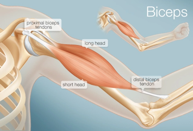
Learn about these muscles, their origin and insertion points, and their functional anatomy. Medical illustration of human arm muscles, veins and nerves. For the formation of a joint, bones need to be joined end to end and fastened with a flexible structure that can allow for movements. / wrist tendonitis, then, is the inflammation of the tendons in the wrist. The tendon that attaches the biceps muscle to the forearm bones (radius and ulna) is called the distal biceps tendon.
Achilles Tendon Disorders - Foot Health Facts from www.foothealthfacts.org Tendons transmit the mechanical force of muscle contraction to the bones. Tendons are thick bands of tissue that connect muscles to bones. Tendon, tissue that attaches a muscle to other body parts, usually bones. Ab 50€ portofrei, versand innerhalb 24h, 100 tage retoure, über 1 mio. Top 10 functions of ligaments forming joints: The achilles tendon is also called the calcaneal tendon. The achilles tendon is the largest. Allows the foot to be turned inward and also supports the arch of the foot.
Its muscle belly is in the forearm.
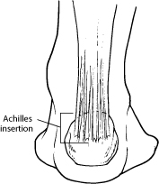
It originates at the back of the femur (thighbone) and patella (kneecap). It runs down the back of the lower leg and connects the calf muscle to the heel bone. Learn about these muscles, their origin and insertion points, and their functional anatomy. A tendon is a band of tissue that connects a muscle to a bone. Below is a diagram of the hamstring tendon. Pin on custom made orthotics. The foot incorporates countless muscles, bones, tendons and ligaments into simple motion and this chart covers them all. Ab 50€ portofrei, versand innerhalb 24h, 100 tage retoure, über 1 mio. Tendons attach muscles to bones. Top 10 functions of ligaments forming joints: The long head of biceps (lhb) is a very important tendon that travels through the shoulder joint (glenohumeral joint).the biceps tendon begins at the top of the shoulder socket (the glenoid) and then passes across the front of the shoulder to connect to the biceps muscle. This muscle diagram is interactive: The knee joint is a complex structure that involves bones.
It can be used by a teacher or student for academic purposes. The fcu tendon is one of two tendons that bend the wrist. You can see how the hamstring muscle connects to the knee via the hamstring tendon on the outside of the knee. Foot anatomy diagram, foot joint diagram, foot sprain diagram, foot tendons and ligaments pain, leg tendon diagram, peroneal tendonitis, foot, foot anatomy diagram, foot joint diagram, foot sprain diagram, foot tendons and ligaments pain, leg tendon diagram, peroneal tendonitis. The tendon that attaches the biceps muscle to the forearm bones (radius and ulna) is called the distal biceps tendon.
The Biceps (Human Anatomy): Function, Diagram, Conditions ... from img.webmd.com Learn about these muscles, their origin and insertion points, and their functional anatomy. The pubis, ischium, and ilium together constitute the pelvis while the thigh bone is the femur. Tendons are similar to ligaments; It originates at the back of the femur (thighbone) and patella (kneecap). They are attached to the femur (thighbone), tibia (shinbone), and fibula (calf bone) by fibrous tissues called ligaments. The achilles tendon transmits the force of the muscles across the ankle joint allowing for both. Tendon, tissue that attaches a muscle to other body parts, usually bones. Tendon diagram simple / 8.4c:
Tendons are found throughout the body, from the head and neck all the way down to the feet.
They are attached to the femur (thighbone), tibia (shinbone), and fibula (calf bone) by fibrous tissues called ligaments. This muscle diagram is interactive: If you feel the outside of your knee you'll feel this tendon. / wrist tendonitis, then, is the inflammation of the tendons in the wrist. Top 10 functions of ligaments forming joints: Learn about these muscles, their origin and insertion points, and their functional anatomy. Ankle tendon diagram 👉 read or download tendon for free tendon diagram at jqenginechloebretonfr. It originates at the back of the femur (thighbone) and patella (kneecap). The bones of the hip include the femur, the ilium, the ischium, and the pubis. Pin on custom made orthotics. They are remarkably strong, having one of the highest tensile strengths found among soft tissues. Joints allow different parts of your body (for example, limbs) to move in different directions. It runs down the back of the lower leg and connects the calf muscle to the heel bone.
Medical illustration of human arm muscles, veins and nerves. The hip itself is a ball and socket joint, much like the shoulder.the structures necessary to create this joint are the socket, the joint capsule, muscle, ligaments, and the neck. The bones of the hip include the femur, the ilium, the ischium, and the pubis. Learn about the anatomy and physiology of tendons. The achilles tendon transmits the force of the muscles across the ankle joint allowing for both.
Quadriceps Tendonitis Information & Treatment Advice ... from www.itendonitis.com The achilles tendon transmits the force of the muscles across the ankle joint allowing for both. Biceps tendons the biceps muscle has two tendons at the shoulder, called the long head and short head. The bones of the hip include the femur, the ilium, the ischium, and the pubis. The two peroneal tendons in the foot run side by side behind the outer ankle bone. One peroneal tendon attaches to the outer part of the midfoot, while the other tendon runs under the foot and attaches near the inside of the arch. If you would like to learn all the parts of the foot structure, you have come to the right place. Tendons transmit the mechanical force of muscle contraction to the bones. The long head of biceps (lhb) is a very important tendon that travels through the shoulder joint (glenohumeral joint).the biceps tendon begins at the top of the shoulder socket (the glenoid) and then passes across the front of the shoulder to connect to the biceps muscle.
Ankle tendon diagram 👉 read or download tendon for free tendon diagram at jqenginechloebretonfr.
If you feel the outside of your knee you'll feel this tendon. It attaches to the wrist bone, the pisiform, and as well as the 5th hand bone. The achilles tendon is also called the calcaneal tendon. Below is a diagram of the hamstring tendon. This muscle diagram is interactive: You can see how the hamstring muscle connects to the knee via the hamstring tendon on the outside of the knee. Hand a hand is a prehensile multi fingered appendage located at the end of the forearm or forelimb of primates such as humans chimpanzees monkeys and lemurs human anatomy for the artist the dorsal hand the dorsal the easiest tendons to identify in the dorsal hand are those of the extensor digitorum muscle its name means extensor of the digits which is The knee is a complex joint that flexes, extends, and twists slightly from side to side. Upper body muscle chart 12 photos of the upper body muscle chart upper body muscle chart, upper body muscle diagram, upper body muscle groups diagram, human muscles, upper body muscle chart, upper body muscle diagram, upper body muscle groups diagram. The largest of these shoulder muscles is the. The tendon connects muscle to the bone. Tendons are found throughout the body, from the head and neck all the way down to the feet. 9 photos of the foot tendons and ligaments diagram.


0 Komentar